Anatomy & Physiology 243

Tom Nordman A&P 243 7:30 Winter 05

Tuesday, March 15, 2005


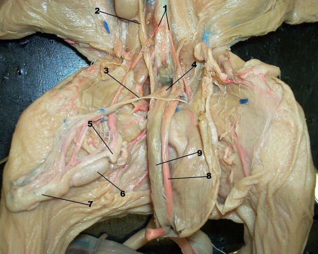
Testis: 1). Epididymis 2). Ductus epididtymis 3). Testis, this whole area 4). Seminiferous tubules 5). Tunica albuginea, this goes on to surround the testis. The germinal epithelium is the wall of each individual seminiferous tubule.
Tom


Testis: 1). Spermatogenic cells 2). Spermatozoa or sperm cells, these will have the flagella (long tails), if you look they are visible in this slide.
Tom


Ovary: 1). Oocyte, w/nucleas 2). Zona pellucida 3). Follicular cavity or antrum, filled w/fluid 4).Granulosa cells/Corona radiata 5). Graafian follicle
Tom


Kidney, injected: 1). Glomeruli
Tom


Ureter: 1).Transitional epithelium 2). Lamina propia 3). Muscularis
Tom

Monday, March 14, 2005


1. Renal pyramid 2.Minor calyx 3.Renal pelvis
Tom


1. Testicular areteries 2.Ureter 3.Vas deferens 4. Seminal vesicle 5. Epididymis 6. Testis 7.Gubernaculum 8. Umbilical artery 9.Bladder
Tom


1.Uretha 2.Urinary bladder 3.Vagina 4. Uterus 5. Ovary 6.Ureter 7. Fallopian tube 8.Broad ligament 9.Urogenital sinus 10. Genital papilla
Tom

Sunday, February 27, 2005

Thursday, February 24, 2005


Lobes of the liver (correct this time)
Tom

If your having troubles viewing all of the photos hit the refresh tab and they will all be there.


another hard to find item
Tom


Looks kind of messy in there.
Tom


The spleen is not on our list, but is good for orientation.
Tom


And who could forget the pancrease
Tom


The blue blob is the kidney, for next section.
Tom


You need to know the anatomy of where these are as they were hard to get good shots of.
Tom


The mesentery is pink
Tom


Papilla is the red dot inside the circle
Tom


Papilla is the red dot inside the circle
Tom


Stomach
Tom


Inside of the stomach
Tom

Saturday, February 05, 2005

Lymphatic histology

These are some of the slides we have been looking at in the lab. I am missing the Trachea, but believe the rest are all here. This site has alot of photos on it so it does not always show all of them, hit your refresh button as soon as you open it up and they will all be available. Another cool thing is you can left click 1 time on any slide and it will show up full screen, hit the back button when done to return to the page. Have a good weekend, Tom
Thanks
Tom


This is a great view of all the different lobes of the fetal pig lungs.
Tom


Fetal pig w/ cricoid and thyroid cartilages, the trachea is also visible here with the C-rings
Tom


Here is a colored drawing of the pig lung, sorry about the quality, I was never to handy with a coloring book.
Tom

Thursday, February 03, 2005


Aveoli
Tom


This is another good example of the red blood cells inside of the capiliaries.
Tom


Hereare a few aveolar sacs inside of a duct.
Tom


The red blood cells are here, you can notice them easy by their color and they are anucleus as well. The blood vessels are another esay find if you look for a structure with the different layers we learned about.
Tom

Wednesday, February 02, 2005


Spleen showing the venous sinuses, the central artery which can be identified by the lumen in the center, and of course we cannot forget our friends the lymphocytes.
Tom


Here is a slide of the spleen w/splenic nodule, germinal center. The red pulp is surrounding the splenic nodules, and the white pulp is in the nodule collectively. Nodules=white pulp.
Tom


Lymph node, showing lymphatic nodule and medulla which is the central portion of the lymph node. The cortex is the outer area containing the nodules.
Tom


Lymph node, w/capsule and cortex
Tom


Lymphatic vessel
Tom

Friday, January 14, 2005


Here is a slide with alot of things visible, the pulmonary artery, pulmonary trunk, ductus arteriosus, aortic arch, subclavian artery, left coronary artery,
Tom

Fetal pig, left subclavian


Left subclavian artery. Brachiocephalic artery. Also there is a good view of the Anterior(superior) vena cava
Tom

Fetal pig, jugular external


External jugular vein with the internal jugular just to the left. The internal goes directly into the brain through the jugular foramen in the base of the skull by the foramen magnum as we learned in 242.
Tom

Fetal pig, common corotid, thyroid


Common left carotid artery. If you jump over the thyroid gland you will have the right one as well.
Tom

fetal pig, posterior vena cava


Posterior (inferior) vena cava.
Tom

Fetal pig, hemiazygos, asending aorta


The white artery is the aorta and the blue vein crossing it is the hemiazygos (azygou)
Tom

fetal pig, aortic arch


Aortic arch.
Tom

Fetal pig, ductus arteriosus


Ductus arteriosus
Tom

Fetal pig, brachiocephalic artery


Brachiocephalic artery
Tom

Fetal pig, left subclavian artery


Left subclavian artery.
Tom

Fetal pig, pulmonary artery


Pulmonary artery
Tom

Fetal pig, left coronary artery


Left coronary artery.
Tom

Fetal pig, thyroid gland


Thyroid gland is located below the larynx and undeneath the thymus glands
Tom

Fetal pig, thymus gland


Thymus gland, this is located on both sides and below the larynx
Tom

Thursday, January 13, 2005

Blood vessels histology


1.)arterial blood 2.)tunica media 3.) lumen of the artery 4.)tunic externa 5.)tunic intima 6.)venous blood 7.)wall of a vein 8.)lumen of a vein 9.)adipose tissue. We took a look at these in the lab on thursday.
Tom

Neutrophil


Neutrophil, this is the most common of the white blood cells 50-70%
Tom

Monocyte


Monocyte, these account for 2-8% of all white blood cells
Tom

Latest news Today I finally posted the histology slides for this section, they are all here except for the bladderwhich will be here tomarrow afternoon. The fetal pig slides are here as well, both female/male and the kidney. Dave took a look at all of them and everything is correct, but it never hurts to make sure. This is the last lab test we will take in A&P, it will be a sad day to leave it all behind but time to move on. Take it easy,Tom email: ibnspokane@comcast.net

Click on the LINKS below

lecture objectives for chapter 25,24,20,19,18.

Digestion quiz

My Photo
Name:
Location: Spokane, Washington, United States

tom

Feel free to give me any feedback on things you might like to see here. ibnspokane@comcast.net