posted by Mickey Mouse @ 6:43 PM
2 comments
![]()
![]()
Tom Nordman A&P 243 7:30 Winter 05
If your having troubles viewing all of the photos hit the refresh tab and they will all be there.
posted by Mickey Mouse @ 2:46 PM
0 comments
![]()
![]()

The spleen is not on our list, but is good for orientation.
Tom
posted by Mickey Mouse @ 2:02 PM
0 comments
![]()
![]()

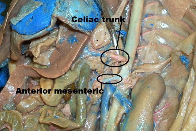
You need to know the anatomy of where these are as they were hard to get good shots of.
Tom
posted by Mickey Mouse @ 1:58 PM
0 comments
![]()
![]()
These are some of the slides we have been looking at in the lab. I am missing the Trachea, but believe the rest are all here. This site has alot of photos on it so it does not always show all of them, hit your refresh button as soon as you open it up and they will all be available. Another cool thing is you can left click 1 time on any slide and it will show up full screen, hit the back button when done to return to the page. Have a good weekend, Tom
Thanks
Tom
posted by Mickey Mouse @ 10:05 PM
0 comments
![]()
![]()

This is a great view of all the different lobes of the fetal pig lungs.
Tom
posted by Mickey Mouse @ 9:59 PM
0 comments
![]()
![]()

Fetal pig w/ cricoid and thyroid cartilages, the trachea is also visible here with the C-rings
Tom
posted by Mickey Mouse @ 9:56 PM
0 comments
![]()
![]()

Here is a colored drawing of the pig lung, sorry about the quality, I was never to handy with a coloring book.
Tom
posted by Mickey Mouse @ 8:48 PM
0 comments
![]()
![]()

This is another good example of the red blood cells inside of the capiliaries.
Tom
posted by Mickey Mouse @ 11:18 PM
0 comments
![]()
![]()

The red blood cells are here, you can notice them easy by their color and they are anucleus as well. The blood vessels are another esay find if you look for a structure with the different layers we learned about.
Tom
posted by Mickey Mouse @ 11:15 PM
0 comments
![]()
![]()

Spleen showing the venous sinuses, the central artery which can be identified by the lumen in the center, and of course we cannot forget our friends the lymphocytes.
Tom
posted by Mickey Mouse @ 7:57 PM
0 comments
![]()
![]()

Here is a slide of the spleen w/splenic nodule, germinal center. The red pulp is surrounding the splenic nodules, and the white pulp is in the nodule collectively. Nodules=white pulp.
Tom
posted by Mickey Mouse @ 7:54 PM
0 comments
![]()
![]()

Lymph node, showing lymphatic nodule and medulla which is the central portion of the lymph node. The cortex is the outer area containing the nodules.
Tom
posted by Mickey Mouse @ 7:51 PM
0 comments
![]()
![]()
 Today I finally posted the histology slides for this section, they are all here except for the bladderwhich will be here tomarrow afternoon. The fetal pig slides are here as well, both female/male and the kidney. Dave took a look at all of them and everything is correct, but it never hurts to make sure. This is the last lab test we will take in A&P, it will be a sad day to leave it all behind but time to move on. Take it easy,Tom email: ibnspokane@comcast.net