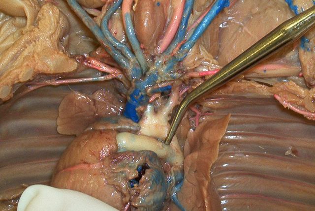
Here is a slide with alot of things visible, the pulmonary artery, pulmonary trunk, ductus arteriosus, aortic arch, subclavian artery, left coronary artery,
Tom
posted by Mickey Mouse @ 6:04 PM
2 comments
![]()
![]()
Tom Nordman A&P 243 7:30 Winter 05
Here is a slide with alot of things visible, the pulmonary artery, pulmonary trunk, ductus arteriosus, aortic arch, subclavian artery, left coronary artery,
Tom
posted by Mickey Mouse @ 6:04 PM
2 comments
![]()
![]()

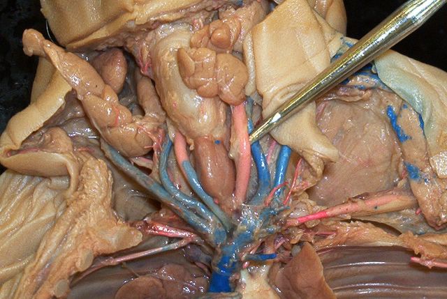
Left subclavian artery. Brachiocephalic artery. Also there is a good view of the Anterior(superior) vena cava
Tom
posted by Mickey Mouse @ 5:58 PM
2 comments
![]()
![]()

External jugular vein with the internal jugular just to the left. The internal goes directly into the brain through the jugular foramen in the base of the skull by the foramen magnum as we learned in 242.
Tom
posted by Mickey Mouse @ 5:49 PM
0 comments
![]()
![]()
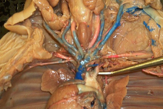
Common left carotid artery. If you jump over the thyroid gland you will have the right one as well.
Tom
posted by Mickey Mouse @ 5:46 PM
0 comments
![]()
![]()

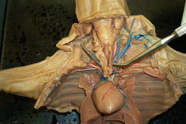
Posterior (inferior) vena cava.
Tom
posted by Mickey Mouse @ 5:43 PM
0 comments
![]()
![]()

The white artery is the aorta and the blue vein crossing it is the hemiazygos (azygou)
Tom
posted by Mickey Mouse @ 5:40 PM
0 comments
![]()
![]()

Brachiocephalic artery
Tom
posted by Mickey Mouse @ 5:33 PM
0 comments
![]()
![]()
Left subclavian artery.
Tom
posted by Mickey Mouse @ 5:32 PM
0 comments
![]()
![]()

Left coronary artery.
Tom
posted by Mickey Mouse @ 5:13 PM
0 comments
![]()
![]()

Thyroid gland is located below the larynx and undeneath the thymus glands
Tom
posted by Mickey Mouse @ 5:02 PM
1 comments
![]()
![]()
Thymus gland, this is located on both sides and below the larynx
Tom
posted by Mickey Mouse @ 4:59 PM
0 comments
![]()
![]()

1.)arterial blood 2.)tunica media 3.) lumen of the artery 4.)tunic externa 5.)tunic intima 6.)venous blood 7.)wall of a vein 8.)lumen of a vein 9.)adipose tissue. We took a look at these in the lab on thursday.
Tom
posted by Mickey Mouse @ 11:59 PM
0 comments
![]()
![]()
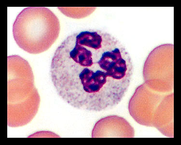
Neutrophil, this is the most common of the white blood cells 50-70%
Tom
posted by Mickey Mouse @ 11:52 PM
0 comments
![]()
![]()

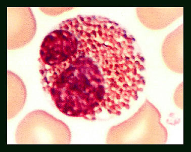
Monocyte, these account for 2-8% of all white blood cells
Tom
posted by Mickey Mouse @ 11:51 PM
0 comments
![]()
![]()

Lymphocyte, these account for 20-30% of all white blood cells
Tom
posted by Mickey Mouse @ 11:49 PM
0 comments
![]()
![]()
Eosinophil, these account for 2-4% of all white blood cells
Tom
posted by Mickey Mouse @ 11:48 PM
0 comments
![]()
![]()

Basophil, these make up less than 1% of all white blood cells
Tom
posted by Mickey Mouse @ 11:45 PM
0 comments
![]()
![]()

This slide of an artery here shows the tunica externa (fiberous connective tissue), tunica media (smooth muscle), tunica interna (endothelium), lumen (hollow space filled with blood). This was done in the 11:30 lab thursday.
Tom
posted by Mickey Mouse @ 6:47 PM
0 comments
![]()
![]()

The pointer is in the middle of a lumen that is filled with blood, this is an artery (round shape). The irregular shaped white spots are veins (flattened or non-circular). If you look carefully you will see the different shades of red surrounding the artery, these are the tunic layers
Tom
posted by Mickey Mouse @ 6:40 PM
0 comments
![]()
![]()

Pancreas showing the Interlobular ducts
Tom
posted by Mickey Mouse @ 6:23 PM
0 comments
![]()
![]()

Adrenal Gland, this is a good example of the Zona glomerulosa and the Zona Fisciculata. The white spot on the middle left is a medullary vein which is found in the Medulla.
Tom
posted by Mickey Mouse @ 6:20 PM
0 comments
![]()
![]()
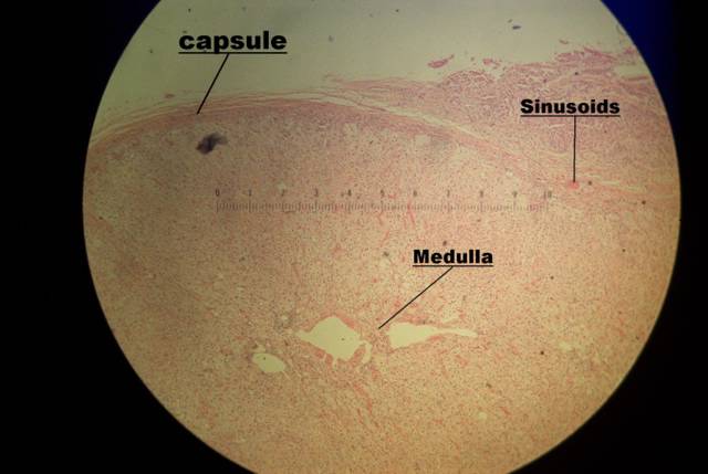
Adrenal Gland.This was a difficult one for me to find a good example of all the points of interest. The capsule is the darkest red line on top.
Tom
posted by Mickey Mouse @ 6:15 PM
0 comments
![]()
![]()
Thyroid Gland in all of it's beauty. I posted this photo for the start of the thyroid series. You can see everything from here but not as clear as the other slides. I put this one in because I think it is a cool photo of a cell, it kinda looks like a planet to me.
Me
posted by Mickey Mouse @ 7:33 PM
0 comments
![]()
![]()

Thyroid Gland showing the follicles, follicular cells, and the colloid in lumen. I checked on the diffinition of "Lumen" and it is just a space. That space is filled with some kind of jelly stuff and that is the Colloid, it has a red color in this slide.
Me
posted by Mickey Mouse @ 7:30 PM
0 comments
![]()
![]()

Thyroid gland, The blue dots here are the nucleas of each follicular (principle) cell, when you are in the lab you will notice the cell membrane around each dot which depicts its size. The parafollicular cells will be the same as these but a bit larger, and also will not come in contact with the follicle directly, but rather, will be surrounded by the smaller follicular cell. Take a look at the Endo lab to view a parafollicular cell.
Me
posted by Mickey Mouse @ 7:25 PM
0 comments
![]()
![]()
Thyroid Gland, Here is a good photo of this gland, #1 is showing the Follicular (principal) cells, these are located around every Follicle and are only a few cells thick being either squamous,cuboidal, or columnar shaped. The dark dots you see are the nucleas of every follicular cell. #2 is showing the Colloid, this appears to me to just some kind of "Goo" which fills the lumen, which is just the space.
Me
posted by Mickey Mouse @ 7:12 PM
0 comments
![]()
![]()
Here is a link to chapter 18 objectives
click here
posted by Mickey Mouse @ 1:09 AM
0 comments
![]()
![]()
Well here we go. I spent the afternoon in the A&P lab trying to get some good photos of the cells we have been looking at this week. After many attemps I finally got it down with the help of a toilet paper tube (empty of course) some tape and alot of patients, well we can't forget the camera, and the cookies I had hidden in my pack. Mainly today I worked on the Thymus gland, and all of the points of interest everyone should know. There will be additions added to this site on a daily basis, even if it is just a message to say I was slacking and no new pics.
posted by Mickey Mouse @ 3:05 PM
2 comments
![]()
![]()

And now for you viewing pleasure, we have a Thymic (Hassall's) Corpuscle that i found while wondering through the medulla today. Take a close look and you will see the Epithelial recticular cells that are in concentric arrangment around the corpuscle at the end of the pointer. (Thymus Gland) If you take a close look at the bottom edge of the slide, you will see where the loose packed cells of the medulla end, and the tightly packed cells of the cortex begin, the colors begin to change here.
Me
posted by Mickey Mouse @ 3:03 PM
0 comments
![]()
![]()

There are 3 Hassall's Corpuscles shown here. 1 to the left of the pointer, 1 to the right, and 1 just above. These Hassall's Corpuscles are located only in the medulla of the Thymus Gland.
Tom
posted by Mickey Mouse @ 2:41 PM
0 comments
![]()
![]()
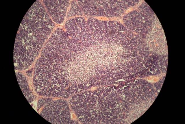
This shows the Cortex, which is darker in color due to the denser arrangement of cells. The cortex is surrounding the Medulla, which is the lighter region in the center of the slide. The red lines are the Thymic Trabeculae which seperate the lobules.
Me
posted by Mickey Mouse @ 2:38 PM
0 comments
![]()
![]()

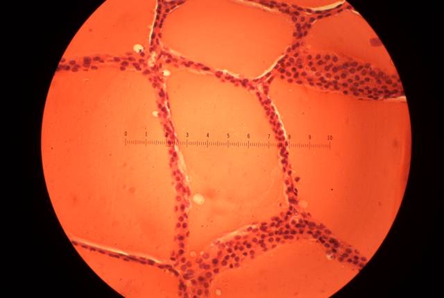
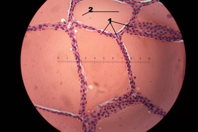
This shows the capsule to the left of the lobules with the interlobular trabeculae seperating the lobules. (Thymus Gland).
Me
posted by Mickey Mouse @ 2:33 PM
0 comments
![]()
![]()
 Today I finally posted the histology slides for this section, they are all here except for the bladderwhich will be here tomarrow afternoon. The fetal pig slides are here as well, both female/male and the kidney. Dave took a look at all of them and everything is correct, but it never hurts to make sure. This is the last lab test we will take in A&P, it will be a sad day to leave it all behind but time to move on. Take it easy,Tom email: ibnspokane@comcast.net