Thyroid gland
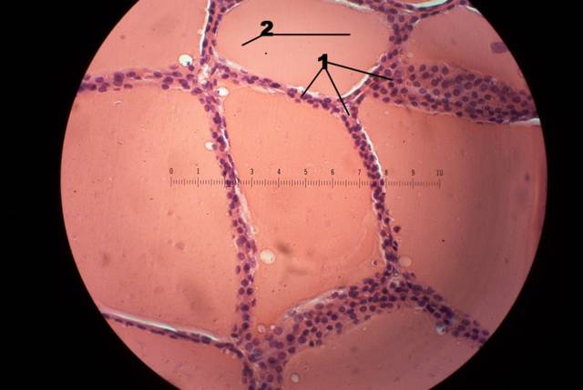
Thyroid Gland, Here is a good photo of this gland, #1 is showing the Follicular (principal) cells, these are located around every Follicle and are only a few cells thick being either squamous,cuboidal, or columnar shaped. The dark dots you see are the nucleas of every follicular cell. #2 is showing the Colloid, this appears to me to just some kind of "Goo" which fills the lumen, which is just the space.
Me
posted by Mickey Mouse @ 7:12 PM
![]()
![]()
 Today I finally posted the histology slides for this section, they are all here except for the bladderwhich will be here tomarrow afternoon. The fetal pig slides are here as well, both female/male and the kidney. Dave took a look at all of them and everything is correct, but it never hurts to make sure. This is the last lab test we will take in A&P, it will be a sad day to leave it all behind but time to move on. Take it easy,Tom email: ibnspokane@comcast.net


0 Comments:
Post a Comment
<< Home